
CASE 5
Kindly provided by Nicky Parry of Tufts Veterinary School.
Clinical history
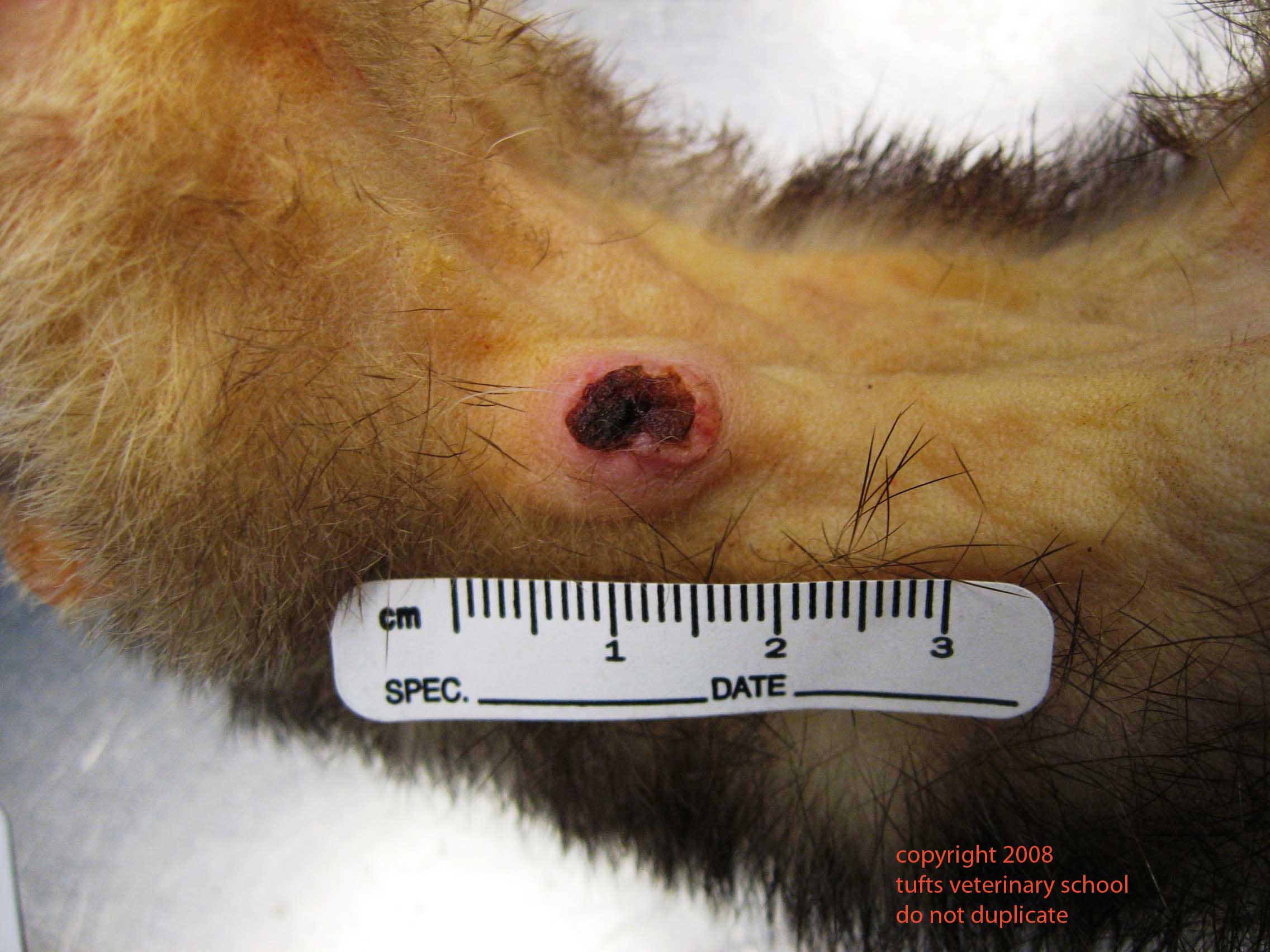
This ventral skin mass was an incidental finding in a three year old female ferret that was presented for postmortem examination.
Describe the gross findings.
Give a morphological diagnosis.
What differential diagnoses might you consider for a skin mass in a ferret?

[expand title=”Reveal Answer”]
Sebaceous epithelioma
Neoplasia is the most common form of skin disease in ferrets, and its incidence increases with age. Although many possible differentials are to be considered, by far the two most common types of tumour seen are mast cells tumours and sebaceous epitheliomas.
Another possibility is a tumour of apocrine origin – these tend to arise mostly in the regions of the head, neck, prepuce, and vulva. They may be benign or malignant but apparently those in the regions of the vulva or prepuce are often malignant.
This particular lesion was a sebaceous epithelioma. These present as wart-like, verrucose masses and although they can arise anywhere on the body, the head and neck regions are predilection sites. These are invariably benign, but may become ulcerated and inflamed. Microscopically they are typically lobulated and comprise proliferations of basal cells (the small blue cells in the photomicrograph) with interspersed sebaceous cells (the larger, foamy-appearing cells).
Parker GA, Picut CA. Histopathologic features and post-surgical sequelae of 57 cutaneous neoplasms in ferrets (Mustela putorius). Vet Pathol (1993) 30:499-504
[/expand]