
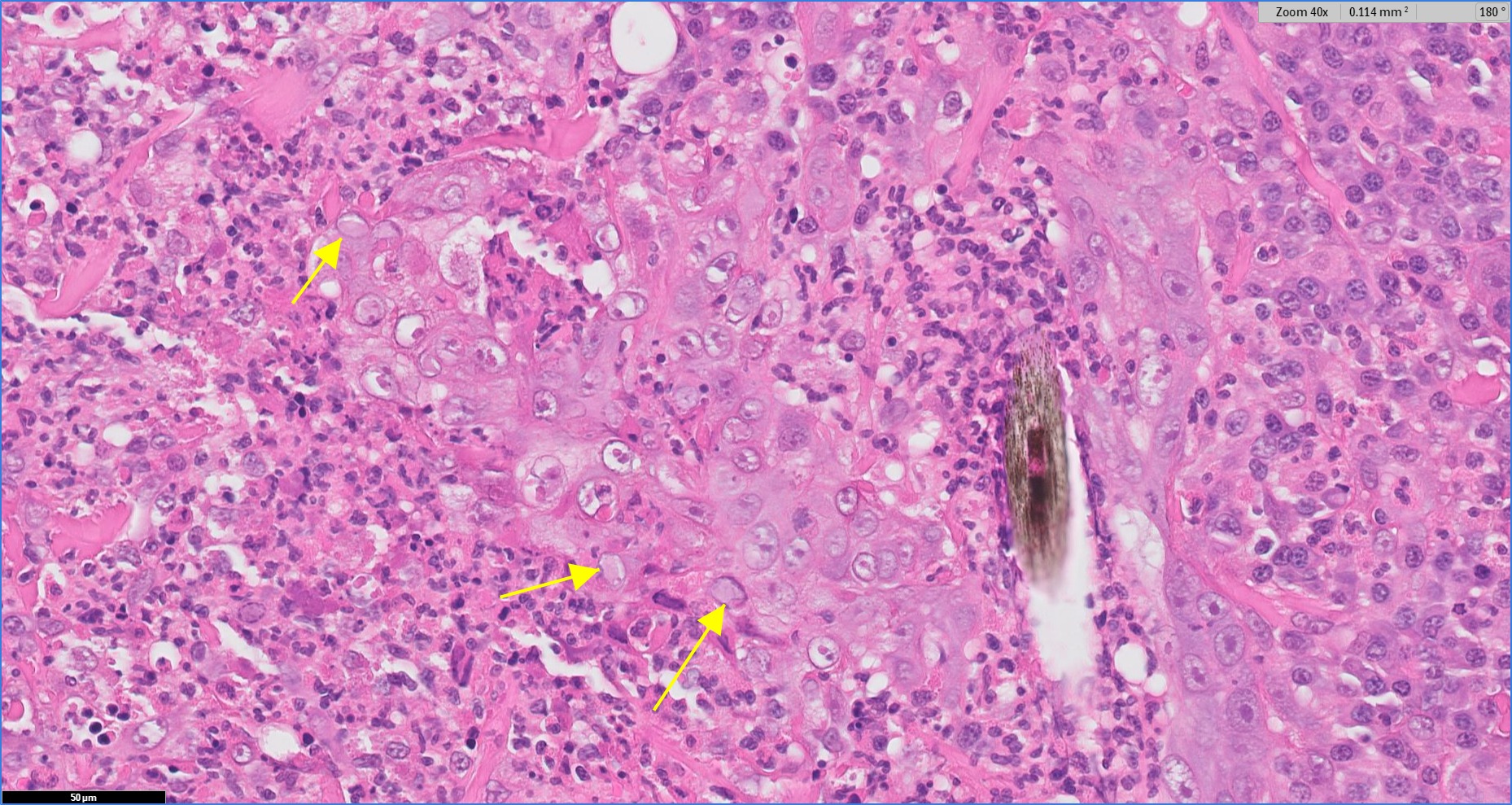
Feline Herpesvirus Infection
Our third and last meeting of the year was held on Wednesday 17th October. We had a strong turn-out with 20 people attending. A brief list of the cases presented is given below.
Nodular Pinnal Dermatopathy in Kittens – Ann Hargis and Jaco van der Lugt of the International Society of Veterinary Dermatology kindly allowed presentation of their cases.
Eosinophilic folliculitis and furunculosis in a dog associated with leptospirosis. – Kate Murphy and David Shearer kindly allowed presentation of their case material.
Leptospirosis in dogs and cats: new challenges from an old bacteria. In Practice (2018) 40 (6) pp 218-229
Isolation of Leptospiral interrogans serovar grippotyphosa from the skin of a dog. JAVMA (1993) 203 (11) pp 1550-1551
Generalized calcinosis cutis associated with probable leptospirosis in a dog. Vet Dermatol. (2005) 16(6) pp 401-6.
Two uncommon manifestations of leptospirosis: Sweet’s syndrome and central nervous system vasculitis. Asian Pac J Trop Med. (2011) 4(1) pp 83-4.
Severe leptospirosis with unusual manifestation. J Trop Pediatr. (2007) 53(1) pp 55-8
Sertoli cell tumour in the scrotum of a castrated dog. And seminoma in the inguinum of a 2 year old castrated cat.
Extratesticular interstitial and Sertoli cell tumours in previously neutered dogs and cats: A report of 17 cases. Can Vet Journal (2006) 47 pp 763-766
Unusual epithelial lined cystic structures in the abdomen of a cat, associated with omentum adjacent to spleen and pancreas. The differentials of mesothelioma, carcinomatosis and duodenal pancreatic heterotopia, particulary Type 3 when mostly ducts are present, were discussed.
https://bmcsurg.biomedcentral.com/articles/10.1186/s12893-017-0250-x
Bilateral optic nerve malacia in a mule. The cause was unknown but trauma is a well recognized cause.
A viral plaque in the cornea of a dog.
Pyogranulomatous opthalmitis associated with Mycobacterium bovis
Concurrent lymphoma and Leishmania onlymph node cytology from a dog.
Neoplastic cells lining the inner aspect of the iris in a dog. The differentials of metastatic carcinoma versus primary iridocilary tumour were discussed.
Osteosarcoma developing in a gossypiboma in a dog.
Synovial lipomatosis in a dog.
And some very nice examples of cutaneous herpesvirus in a cat with some lovely inclusions. A GIST in a cat, cutaneous leishmaniasis in a dog and botryomycosis in the skin of a cat due to Staph. aureus.
A fibrovascular benign cutaneous nodule likely angiofibroma in a dog.
A collision tumour in the urinary bladder of a dog (tcc and hemangiosarcoma).
An odontogenic tumour with squamous differentiation and desmoplasia in a cat – desmoplastic ameloblastoma.
Nodular proliferation of bland epithelioid and spindle cells in the distal limb of a cat (it was affecting subcutis and metacarpal bone tissue), from which a glomus tumor was suspected, but due to some apparent chondroid foci, the possibility of a chondroid tumour was also considered