
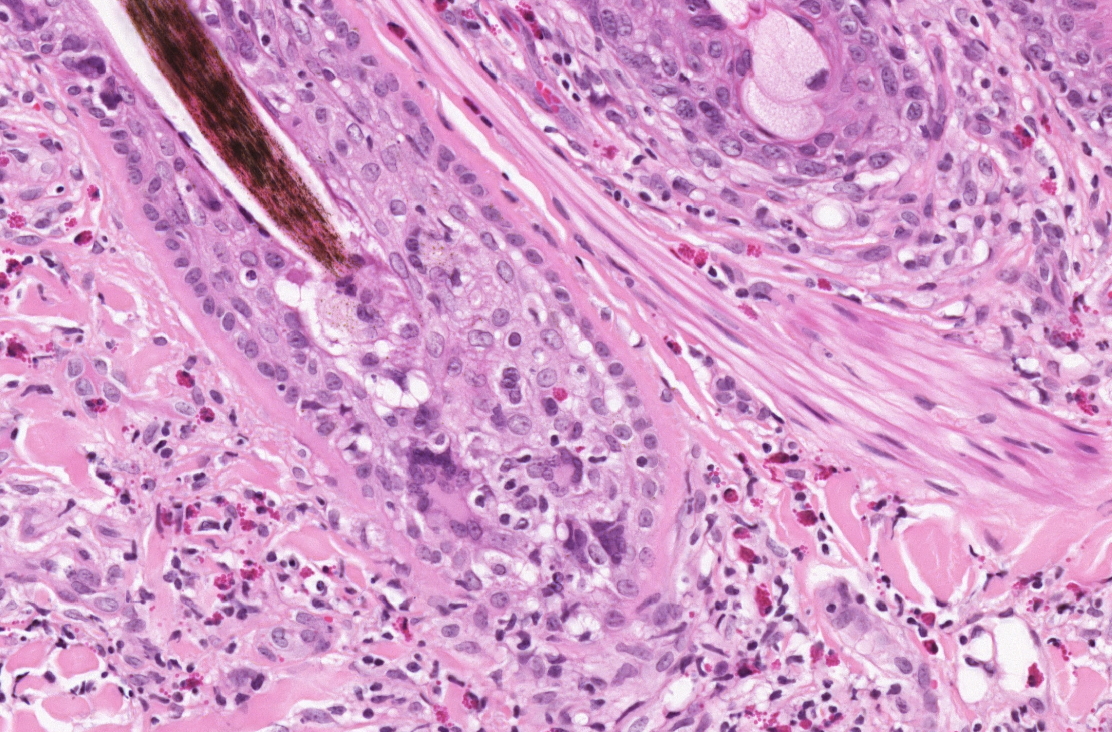
Equine skin: granulomatous mural folliculitis with giant cells.
Our first Bristol Histopath Slide Club meeting of the year was held on the 19th April. As usual there was a wide range of pathology presented. A brief overview of the cases presented is included. We saw a case of Paramyxovirus 1 in a pigeon, myoepithelioma and a unusual presentation of gastric ulceration, oral mucosal ulceration and neutrophilic dermatitis in a mouse. There was an unusual meningeal tumour in a Border Terrier, differentials were discussed but sadly the case could not be investigated further with immunohistochemistry. A case of interface onychitis was presented and the problems of sampling and diagnosis were discussed with this condition or group of conditions that may wax and wane. A good review of interface onychitis is given in the article referenced below. There was a case of granulomatous mural folliculitis, eosinophilic dermatitis and vasculitis involving the limbs only in a horse and the appearance of normal juvenile rabbit skin and Filta-bac cream on H&E section. Sadly the technology failed for the digital slides, or we would have seen a granulomatous mesenteric lymphadenitis in a cat which was acid fast positive and a persistent vitelline duct cyst.
Pathology in Practice: Symmetric lupoid onychodystrophy in a dog. JAVMA (2015) 246(2) 197-199