
Our second Bristol Histopath Slide Club meeting was held on 15th May. A brief overview of the cases submitted follows.
Several cases prompted more extensive discussion. A dermal mass on the ear of a dog was considered to be a giant cell granuloma and the diagnostic difficulties of this entity were discussed. An amazing case of osteonecrosis of the jaw in a dog was presented initiating discussion as to possible pathogenesis. A case of multiple cutaneous mast cell infiltrates in a Puggle prompted discussion of the differential diagnosis of mast cell tumour versus mastocytosis in young dogs.
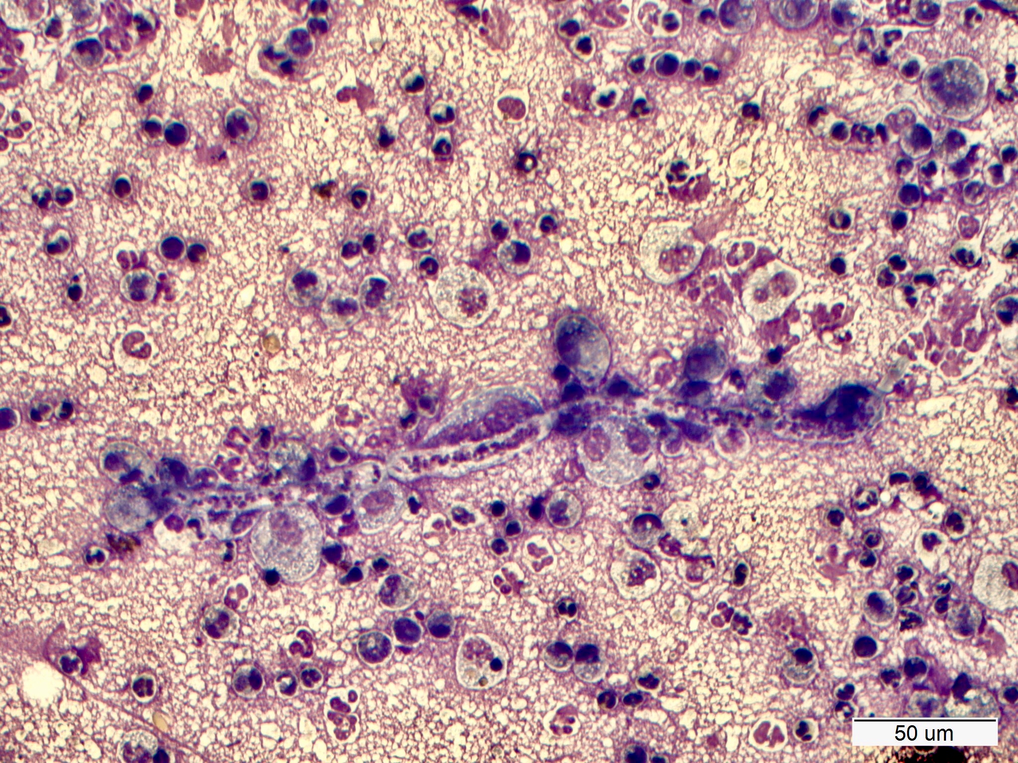
Nice examples of classic pathology included a cowpox virus infection in the skin of a Mara, staphylococcal folliculitis and furunculosis in a horse and a subchondral bone cyst in a horse. We saw cytology of cutaneous dirofilariasis with a microfilaria undergoing phagocytosis (pictured).
Unusual cases included a thymoma in a cross bred dog showing melanisation, a nice case of multiple plasmacytomas in a dog and a vascular hamartoma in the gingiva of a cat.
There was a case of non suppurative encephalitis in a miniature horse with a viral origin the main differential. A further case of encephalitis in a cat, presumed Toxoplasmosis and vegetative valvular endocarditis in a dog with spectacular gross pathology.
References.
Osteonecrosis of the Jaw.
Non-radiation related osteonecrosis of the jaws in dogs: 14 cases (1996-2014) Peralta, S et al, Frontiers in Veterinary Science, May 2015, 2:7
Giant cell granuloma
Clinical, histological and prognostic features of a novel nail-bed lesion of cats: 41 cases. Melanie Dobromylskyj, Rebecca Fernandes, Adrienne French. Journal of Feline Medicine and Surgery (2017) 19(8) pp 853-859
Melanocytes in the thymus
Clinical and pathologic features of thymoma in 15 dogs. Aronsohn MG et al J Am Vet Med Assoc. 1984 Jun 1;184(11):1355-62.